


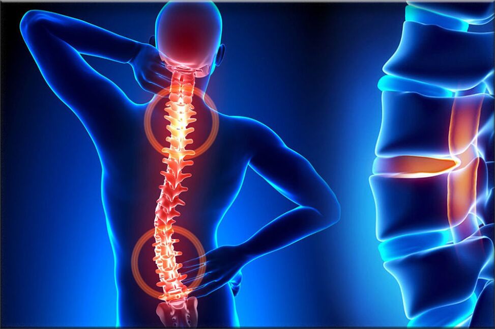
Osteochondrosis is one of the most common diseases of the musculoskeletal system, which manifests itself as a result of a complex of certain dystrophic changes in the cartilage of the spine, during which the lumbar discs are often affected. Structures with intervertebral cartilage discs provide elasticity and at the same time allow the human spine to move, ie provide movement.
With osteochondrosis, a number of processes occur in the vertebral discs that cause degeneration, resulting in a loss of elasticity and a decrease in the degree of elasticity, during which the disc itself becomes very flat. The distance between the two discs is reduced, while at the same time squeezing the nerve endings and blood vessels, causing severe pain. The area of compression of the nerve node begins to swell, which leads to increased pain and even more disruption.
During the development of osteochondrosis, muscle structures and most organs of the body are often involved in this pathological process. This is due to the disruption of blood circulation and mobility of muscles and organs during the maximum disruption of the vascular bundle. For example, the most common osteochondrosis is cervical osteochondrosis, which is accompanied by pain in the back of the head, nausea, dizziness, visual disturbances, and often tinnitus. The disease has become quite "rejuvenated": a century ago, osteochondrosis was a disease of people of gerontological age, and today young people are susceptible to it.

The most vulnerable category of people are people with disorders of the vascular and venous nature, as well as severe disorders of metabolism and hormonal levels in the body. The reason for this is that these diseases lead to impaired oxygenation of the disc. If qualified, timely measures are not taken to heal, the edges of the compressed intervertebral disc will anatomically extend beyond the boundaries of the spine, thereby destroying the nerve bundles.
Therefore, the patient is at risk of getting a herniated disc. The main, important cause of osteochondrosis is the uneven distribution of the spine, which leads to changes in the cartilage structure at points with excessive pressure. The nature of this disease depends on the stage and level of damage to the affected discs. Intervertebral discs change with age, like our hair. Major injuries or fractures of the spine can affect their functioning. Casual wear and certain types of vibration can also accelerate the rate of spinal degeneration. In addition, evidence suggests that smoking increases the rate of spinal degeneration. Scientists have found a link between family members and stressed the role of genetics in how quickly change occurs.
The disease can be caused by various factors:
- injuries, bruises;
- dystrophy of the spinal muscles;
- curvature and curvature of the spine;
- lift weights;
- staying in one position for a long time;
- metabolic disease;
- trace elements and vitamin deficiency - manganese, magnesium, zinc and vitamins D and F;
- hereditary predisposition;
- physical overload;
- sedentary lifestyle;
- radiation background;
- donma;
- congenital dystrophies;
- asymmetric work of the muscles of the spine;
- stress, depression.
These causes of osteochondrosis are only the hypotheses of scientists, they are direct factors that cause the disease, science has not yet found, and we are talking only about risk factors.
The first perioddevelopment - characterized by early placement of the intradiscal nuclear pulposus (nuclear pulposus of the eccentric intervertebral disc near the dorsal part of the vertebrae).
The second periodcharacterized by the appearance of instability of the spinal segment. Pathological substrates are represented by degenerative processes of ascent and posterior longitudinal ligament fragmentation with the fibrous nucleus of the affected disc, pathological movements between the vertebrae develop.
The third perioddevelopment of the disease - general damage to the intervertebral disc, with the appearance of a "herniated disc" - the appearance and appearance of fragments of the pulposus nucleus outside the intervertebral space.
If the disease has reached the third stage, the process of destruction is already irreversible and can lead to deep disability.
Types of osteochondrosis
The evolution of osteochondrosis is slow, spinal injuries, sports, weight lifting, etc. The clinic depends on the location of the lesion.
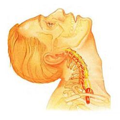
Osteochondrosis of the cervical spinehas local and distant symptoms of advanced forms - with high root dominance, ie contributes to the development of severe radicular pain. Symptoms of osteochondrosis of the cervical spine are accompanied by varying degrees of dysfunction, sometimes manifested by a sudden restriction of mobility of the cervical spine and functional blocks. Headaches can be both traction and paroxysmal, with radiation to the intercapular region or shoulder area. In the acute period, patients are diagnosed with pain attacks in the neck that obstruct and restrict the movement of the head and neck. In addition to severe anxiety, pain syndrome may be accompanied by dizziness, insomnia, pain, loss of appetite, depression, and eye and pharyngeal disorders.
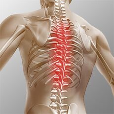
Chest osteochondrosis. . . Clinical manifestations are associated with local lesions and the destruction of the root structure of the nerve. Thoracic osteochondrosis is a pain syndrome of chronic or acute low back pain with chest discomfort and limited muscle contracture, ranging from muscle atrophy in the right mouth. Chest pain can present as diffuse, intercostal, and neuralgic. Palpation increases the axial rotation of the vertebral body. The disorders correspond to the level of root irritation from Thl1 to Thl2 and may manifest themselves as angina pectoris, which is reflected in disorders of the liver and gastrointestinal tract. Disorders of the genitourinary system and genitals often occur. Patients report a significant reduction in sensory disturbances, such as paresthesia, superficial and deep sensitivity.
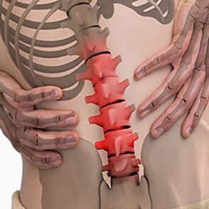
Lumbar osteochondrosis. . . It is characterized by abdominal reflexes and dysfunction of the lower extremities. During the development of neurological diseases, muscle weakness in the legs and dysfunction of the pelvic organs may occur. Osteochondrosis is characterized by an assessment of the damage to the sitting process. The more advanced the stage of spinal cord injury, the shorter the time a patient can sit. Lumbar forms are characterized by chronic and acute low back pain, spasm of the paravertebral muscles, and myofascial secondary syndrome. The pain spreads to the thigh and posterior ilium.
Depending on the localization of the pathological process of osteochondrosis, the disease can lead to a violation of the patient's superficial sensitivity (tactile, thermal). Changes in reflexes (eg, no Achilles reflex), muscle shedding, impaired muscle tone, autonomic disorders (pallor, redness of the skin, trophic changes in the nails, hypothermia of the skin in the distal extremities), sphincter dysfunction are also characteristic. and sexual disorders.
Clinical picture
DiagnosticsIt starts with a complete history and physical examination. The doctor asks questions about the symptoms and how the disease interferes with the patient's daily activities. In addition, the specialist is interested in identifying positions and activities that emphasize or reduce the level of pain.
The doctor then examines the patient, examines the condition of the spine and the degree of movement, and thus determines which movements cause pain. Skin sensitivity, muscle strength and reflexes are checked equally. Based on the medical history and physical examination, the doctor will determine which techniques will help.
Radiography rarely helps in diagnosis, with more than 30% of radiographic images showing abnormalities in the early stages of the disease.
However, if the symptoms are severe and the disease is already in the second or third stage, the image may show defects in one or more intervertebral discs. It can be penetrated by osteophytes between the spine and joints.
If additional information is needed, magnetic resonance imaging is assigned. MRI is used to look at the soft tissues of the body. This is useful if the tissue nucleus absorbs water or if there are cracks inside the disc. MRI can show problems in other soft tissues, such as the spinal nerves.
Discography can help with diagnosis. This test is performed with a contrast agent injected into one or more discs, respectively. Further examination in radiography provides useful information about the condition of the discs.
Treatment of osteochondrosis depending on the variety
Non-surgical treatment of osteochondrosis
Whenever possible, doctors prefer non-surgical treatment. The most important thing in non-surgical treatment is to eliminate pain and other discomforts so that the patient can maintain a comfortable life as much as possible.
Doctors rarely prescribe bed rest to patients with osteochondrosis. Patients are encouraged to live in natural mobility when the pain is not bothersome. If the symptoms are severe, a few days of bed rest may be prescribed.
When changing the location of the spine, an elastic band is sometimes worn for more than 2-4 days to avoid atrophy of the back muscles.
Osteopathic sessions provide serious relief from osteochondrosis.Osteopathic doctornot only diagnoses a problem area, but also relieves pain in 1-2 doses, eliminates the general condition of the body and "squeezes" the visceral organs.
Patients may be prescribed medication to control symptoms and resume normal functioning for a long time. If symptoms continue to limit the patient's activity, a regular doctor may suggest an epidural steroid injection.
Steroids are powerful anti-inflammatory drugs that help relieve pain and inflammation. Non-steroidal anti-inflammatory injections are injected into the space around the spinal roots of the spine. This area is called the epidural space. Some doctors inject steroids alone. However, it is often combined with other drugs. Basically, steroids are prescribed only when other drugs are ineffective, but osteopathy almost always helps.
In addition, patients often work with physical therapists. After assessing the patient's condition, the therapist prescribes exercises to reduce symptoms. The exercise program aims to improve flexibility and is useful for training the abdominal and lumbar muscles to allow them to move with minimal pain.
Surgery
People with osteochondrosis generally do not need surgery. In fact, only 1-3% are in working condition. Surgeons prescribe non-surgical treatment, ie craniosacral osteopathy, as a rehabilitation treatment for at least 3 months before surgery. If there are no results after 3 months of non-surgical treatment, then there are grounds to indicate a surgical procedure.
Basic surgical procedures
Disectomy
The procedure is aimed at partial or complete removal of the disc in the lumbar region. Surgeons usually perform the operation by incision in the lumbar region. Before removing a torn disc, it is necessary to remove part of the boards.
Today, surgery has adopted minimally invasive techniques that require only a small incision in the lumbar region. Proponents of this method claim that it is safe. They also believe that the procedure prevents scars on nerves and joints and helps patients heal faster.
Combine
It is an intervention that connects two or more bones together to prevent wear and tear on the ends of the bones and joints.
Rehabilitation
The doctor may recommend that the patient see a physical therapist several times a week for 4-6 weeks. In some cases, patients need additional help.
The first year of treatment is needed to control symptoms. The therapist will work with you to find pain-relieving positions and movements. Heat, cold, ultrasound and electrical stimulation may be prescribed to relieve pain and muscle spasm. Massage or specialized forms of soft tissue mobilization can also be used. These procedures help the patient to perform the movements easily.
Typically, adjusting the treatment helps to restore the sensitivity of the spinal nerves and muscles, reduce pain, and improve mobility.
The main goal of therapy is to teach the patient to manipulate to prevent future problems. A number of actions are recommended to improve patient comfort. The patient will also be given a strategy to help with recurrent symptoms.
Everyone should learn and take into account all types of osteochondrosis to prevent the development of this disease in themselves and their loved ones. After all, it is impossible to treat the destroyed vertebrae, the therapy is to eliminate the symptoms of pain and achieve a long-term remission. You should also remember a simple but effective rule:The best treatment is prevention. . .
Prevention of osteochondrosis
Prevention is quite simple - it's a healthy diet, regular muscle activity, daily morning warm-ups, a healthy and active lifestyle and monthly visits.osteopathic sessionsto correct and relieve musculoskeletal tension. Adherence to these rules is enough to never face the above problem and to avoid terrible symptoms and lifelong treatment.